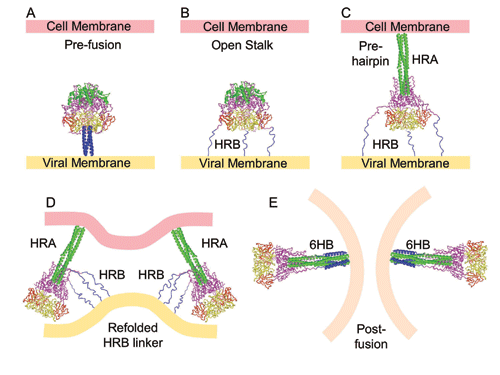
Fig. 7. A model for F-mediated membrane fusion. (a) Structure of the pre-fusion conformation. HRB is colored blue, HRA is colored green, domains I, II and III are colored yellow, red and magenta respectively. (b) An "open stalk" conformation, in which the HRB stalk melts and separates from the pre-fusion head region. HRB is shown as three extended chains because the individual segments are unlikely to be helical. This conformation is consistent with a low-temperature intermediate that is inhibited by HRA peptides, but not HRB peptides. Mutations of the switch peptide residues, 443, 447 and 449 would influence the formation of this intermediate, by affecting stabilizing interactions between the pre-fusion stalk and head domains. (c) A pre-hairpin intermediate can form by refolding of DIII, allowing the formation of the HRA coiled coil and insertion of the fusion peptide into the target cell membrane. This intermediate can be inhibited by peptides derived from both HRA and HRB regions. (d) Prior to forming the final 6HB, the close approach of viral and cellular membranes may be trapped by folding of the HRB linker onto the newly exposed DIII core, with the formation of two b-strands. (e) The formation of the post-fusion 6HB is tightly linked to membrane fusion and pore formation, juxtaposing the membrane interacting fusion peptide and TM domains. (From Yin, H.S., Wen, X., Paterson, R.G., Lamb, R.A. and Jardetzky, T.S. (2006). Nature 439, 38-44. Copyright 2006 by MacMillan Publishers Ltd.)